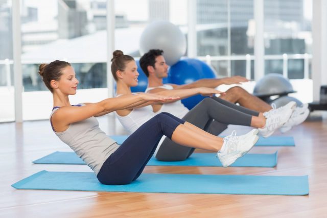
System of muscles
We use our muscles a lot in our daily life—and there are 600 of them in the human body. That is why it’s highly important to learn how to make them stronger and more functional while reducing the risks for injuries.
But if you’re talking about muscle building, conditioning, and strengthening, you can only achieve that with proper weight training and exercise. And one of the best ways to achieve efficiency in resistance training is to learn the basic structure and function of your muscles first.
There are three main muscle groups—skeletal, cardiac and smooth. But in this guide, we’ll focus on the skeletal muscles, which is divided into different muscle groups that most people are familiar with, especially those who love to exercise.
For instance, the pectoral muscles on the chest are responsible for those pressing movements when doing push-ups and bench presses. These large bundles of muscle tissue are connected to the bones by tendons, so their contractions will help facilitate the movement of those bones.
The anatomy of a muscle
Each muscle tissue is composed of bundles of fascicles, which are also composed of bundles of individual muscle fibers. Also called myocytes, these muscle fibers are responsible for movement and each fiber contains multiple nuclei, which increases through the process of protein synthesis that’s stimulated by exercise. This is one of the simplest explanations for how muscles grow.
Muscle cells are filled with sarcoplasm, a type of fluid that facilitates the exchange of nutrients happening within the cells. It also contains glycogen that the muscle tissue uses for energy production and myoglobin where oxygen is stored to help with energy production within the muscles.
Within the cells are myofibrils where muscle contraction takes place. These are composed of sacromeres or long tubes of proteins that are responsible for the muscle contraction that you feel when exercising. Each sarcomere is composed of different strands of protein and each muscle cell can contain anywhere from hundreds to thousands of sacromeres.
Myosin and actin are two of the most important protein strands in sarcomeres because they are attached to each other and are responsible for shortening, which in turn stimulated muscle contraction. During the negative portion or eccentric part of a movement, the myosin filaments release the actin filaments to stimulate the lengthening of the sacromeres. When the muscle is lengthened under stress as with heavy weight training, the action is torn from the myosin. This muscle will eventually repair itself and that is where muscle mass is developed.
The most important muscle groups in exercise
Now that we already have a basic understanding of the anatomy and physiology of the muscle, let’s take a closer look at the different system of muscles that are involved most in fitness training.
The chest
Also known as the pectoralis major, the chest muscle is one of the most prominent muscles in the body and it’s composed of the upper and lower pectoralis major. The pectoralis major is located on the anterior surface of the thoracic cage and many fitness buffs know it as the pecs.
Its main function is for the adductions and internal rotation of the arm. Its movement is stimulated when you perform exercises such as the incline dumbbell press, bench press and push-up.
You can also perform isolation exercises that move the arms without including the elbow joint. These include the dumbbell fly, cable crossover and pec deck.
The chest muscle anatomy:
- Pectoralis major: The largest among the pectoralis muscles, it connects the sternum (breastbone) and clavicle (collarbone) to the humerus (upper arm bone) and anterior deltoid (front of the shoulders).
- Pectoralis minor: This muscle is located beneath the pectoralis major and it runs from the rib cage to the scapula (shoulder blade).
- Serratus anterior: This muscle lies along the side of the rib cage connecting it to the scapula (shoulder blade).
- Subclavius: This muscle is attached to the first rib and clavicle where it forms the front of the axillary muscle (armpit).
The chest muscle physiology:
- Pectoralis major: This muscle is responsible for the flexion of the shoulder joint to allow adduction (inward movement), extension (forward movement) and internal rotation of the humerus.
- Pectoralis minor: This muscle helps to stabilize and depress (downward pull) the scapula.
- Serratus anterior: This muscle pulls the scapula forward when the ark is extended in front of and away from the body.
- Subclavius: This muscle contracts to pull the shoulder downward and forward.
Fitness training tip:
- The different sections of the chest muscles are hit differently depending on the type of chest exercise that you do. So, if you want even results when building muscle, you need to perform exercises that target the upper, middle and lower pectoralis muscles.
Common chest exercises
- Bench press
- Cable crossover
- Dips
- Flye
- Push-up
- Reverse-grip bench press
The back
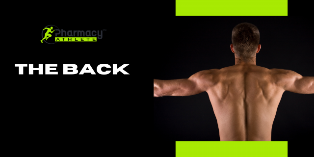
Making up the back part of your torso, the back muscles are mostly composed of the large latissimus dorsi muscles (lats) that run from the upper arms down to the buttocks.
You also have the teres major, the rhomboids and the middle and lower portions of the trapezius muscles. Back muscles can also refer to the musculature of the lower back itself.
These group of muscles are located in the lower back and they support the spinal column. They include the spinal erectors like the longissimus thoracis, spinalis thoracis and iliocostalis lumborum.
The back muscle anatomy:
- Latissimus dorsi: This muscle runs from the spinal vertebra and iliac crest (top of the hip bone) to the humerus (upper arm bone).
- Rhomboids: This muscle lines underneath the trapezius muscles and are attached to the spine and scapula (shoulder blade).
- Teres major: This muscle is located above the lats and connected to the scapula and humerus.
- Teres minor: This is a type of rotator cuff muscle that spans from the scapula to the upper portion of the humerus and the shoulder joint.
- Erector spinae: These are muscle fibers and tendons that originate from the base of the spine and pelvis (hip bone) and runs the length of the back to various ribs and vertebrae.
The back muscle physiology:
- Latissimus dorsi: It is responsible for the extension, vertical adduction (inward movement) and horizontal abduction (outward movement) of the humerus.
- Rhomboids: This muscle helps retract the scapula.
- Teres major: It helps with the extension and internal rotation of the humerus.
- Teres minor: It works with the rear deltoid (shoulder muscle) to externally rotate the humerus.
- Erector spinae: These muscles help with stabilizing, straightening and rotating the back.
Fitness training tip:
- Pulling and rowing exercises are two of the major types of lat exercises that work on the back muscles. They include pull-ups, pulldowns, T-bar rows, bent-over barbell rows and seated cable rows. Pull-up and pulldown exercises target the teres major and outer lats while rowing exercises focus on the middle and lower lats, the middle trapezius muscles and the rhomboids.
Common back exercises
- Barbell rows
- Cable pulldowns
- Good mornings
- Pull-ups
- Seated cable rows
The shoulders
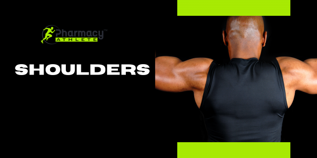
Shoulder muscles are basically the deltoids located on top of the upper arm. Composed of three heads that originate from different points of the shoulder girdle—the anterior, lateral and posterior heads—and converging on one common tendon that inserts to the humerus, the deltoid muscles form the rounded contour of the shoulders and they help prevent the subluxation or dislocation of the head of the humerus, especially when carrying a heavy load.
The shoulder muscle anatomy:
- Anterior deltoid: This muscle begins at the clavicle (collarbone) and the pectoralis major, and converges at the humerus with the other deltoid heads.
- Lateral deltoid: This muscle spans from the acromion of the scapula (shoulder blade) to the same convergence point.
- Posterior deltoid: This muscle attaches to the same convergence point from the spine of the scapula on top of the shoulder blade.
- Rotator cuff: This is composed of the supraspinatus, infraspinatus, subscapularis and teres minor muscles that connect the scapula to the humerus.
The shoulder muscle physiology:
- Anterior deltoid: This works together with the pectoralis major during pressing movements and with the subscapularis during internal humeral rotation.
- Lateral deltoid: This is responsible for abduction and overhead pressing movements.
- Posterior deltoid: This works with the latissimus dorsi to extend the arm backward.
- Rotator cuff: These muscles assist with the rotation of the humerus.
Fitness training tip:
- Since each of these heads are targeted by different exercises, it’s important to work out using basic multi-joint movements that hit all three heads at one time. Isolation exercises like lateral raise, front raise and rear deltoid raise are also great to work out these shoulder muscles.
Common shoulder exercises:
- External rotation
- Face pull
- Internal rotation
- Lateral raise
- Rear delt raise
- Shoulder press
The biceps
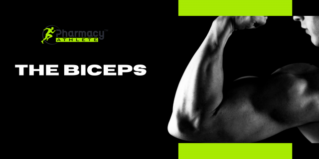
One of the most familiar muscles for fitness enthusiasts is the biceps brachii, which is essentially the muscles on the front part of each upper arm. It is composed of a short head (inner) and long head (outer) that work together as a singular muscle.
The major difference between the two is right where they attach on the scapula. The tendon of the long head muscle attaches farther back on the scapula than the short head, thus the name. Both these muscles join into a single tendon near the elbow, which attaches to the radius to cause flexion of the elbow when the muscles contract.
The biceps anatomy:
- Biceps brachii: Both the long and short head attach to the scapula. The long head originates from the supraglenoid tubercle of the scapula while the short head originates from the coracoid process at the front of the scapula.
- Brachialis: This runs from the middle part of the humerus (upper arm bone) to the ulna (forearm bone).
- Brachioradialis: This connects the lower end of the humerus to the end of the radius near the wrist.
Fitness training tip:
- The biceps get some assistance from the brachialis muscle when flexing the elbow. Lying underneath the biceps muscles, it helps with the first 30 degrees of elbow flexion.
This muscle is also involved with elbow flexion when the hands maintain an overhand grip on the bar. The brachioradialis is also useful in initiating elbow flexion when the hand is in neutral position such as when you’re doing hammer curls.
Common biceps exercises:
- Biceps curl
- Chin up
- Concentration curl
- Hammer curl
- Preacher curl
- Reverse-grip curl
The triceps
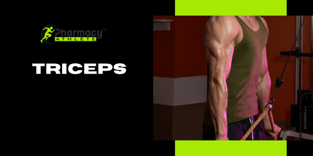
From its name, the triceps brachii muscle is composed of three muscle heads—the lateral, long and medial heads—on the back of the upper arm. Each head is attached differently on the upper end, but they join together into a single tendon that crosses the elbow and attaches to the ulna.
The triceps muscle anatomy:
- Triceps brachii: From their beginning point, the three heads of the triceps converge at the olecranon of the radius or the elbow. Both the lateral and medial heads begin at the humerus while the long head begins at the scapula.
- Anconeus: Lying beneath the triceps brachii, this muscle originates right at the middle of the humerus and ends at the olecranon just like the triceps brachii.
The triceps muscle physiology:
- Triceps brachii: It initiates the extension of the arm at the elbow and it also functions during pressing movements that extend the arm away from the body.
- Anconeus: this assists in the extension of the arm.
Fitness training tips:
- Compound exercises and isolation movements are the two best types of exercises for the triceps muscles. Compound exercises involve the extension at the elbow and movement at the shoulder like what you would do in dips and close-grip bench presses. Isolation exercises, on the other hand, involves the extension at the elbow just like you would with dumbbell kickbacks.
- Although all three triceps heads are targeted in every exercise, the long head is contracted more strongly during exercises because it attaches to the scapula. This is why exercises that are done overhead puts the most stress on the long head of the triceps.
- Exercises that focus on extending the arms at the sides of the torso while holding a neutral or overhand grip target the lateral triceps head while exercises that are done with an underhand grip stresses the medial head.
Common triceps exercises:
- Close-grip bench press
- Dips
- Triceps kickback
- Triceps overhead extension
- Triceps pressdown
The abdominals
Another highly utilized muscle group during workouts are the abdominals, which are four muscles on the midsection of the body named the rectus abdominis, external obliques, internal obliques and transverse abdominis.
The abdominal muscle anatomy:
- Rectus abdominis: This is what you see as the visible abs that run from the front of the pelvis to the ribs and sternum, which is divided by a vertical line of connective tissue called the linea alba.
- Internal and external obliques: These are the side muscles of the abdomen, which originate from parts of the lower ribs and connect to the ilia.
- Transverse abdominis: This lies beneath the rectus abdominis and obliques where it connects the bones of the rib cage to those of the pelvis.
The abdominal muscle physiology:
- Rectus abdominis: It flexes the spine bringing the rib cage toward the pelvis or vice versa.
- Internal and external obliques: It is responsible for stabilizing the spine, compressing the abdomen and the rotation of the trunk and lateral flexion of the spine.
- Tranverse abdominis: It is responsible for stabilization and compression.
Fitness training tips:
- Crunch exercises help target the upper abs while the lower abs are best trained with exercises that involve flexion of the lower spine forward by bringing the knees toward the chest.
- Exercises that flex the spine laterally to the left and right such as the oblique crunches are best for targeting the internal and external obliques. You can also do exercises that involve flexing the spine forward and rotating it to the left or right such as with crossover crunches.
- Core exercises that flex the transverse abdominis pulling the navel in toward the spine is best used for targeting the deep transverse abdominis.
Common abdominal exercises:
- Cable woodchopper
- Crunch
- Hanging leg raise
- Plank
- Reverse crunch
The quadriceps
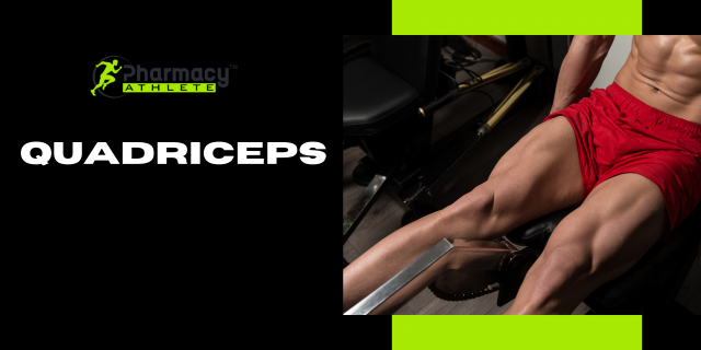
The quadriceps are four muscles—the vastus lateralis, vastus medialis, vastus intermedius and rectus femoris—form the front of the thigh. These muscles have different points of origin on the thigh and hip bone, but they converge on a single tendon to perform knee extension.
The quadriceps muscle anatomy:
- Rectus femoris: It originates at the ilium and connects to the tibia, one of the two bones of the lower leg.
- Vastus lateralis: The outermost part of the three muscles underlying the rectus femoris, it connects the femur to the tibia.
- Vastus intermedius: It is located between the vastus lateralis and vastus medialis and runs from the femur to the tibia.
- Vastus medialis: This is the innermost of the three muscles underlying the rectus femoris and it connects at the femur and tibia.
The quadriceps muscle physiology:
- Rectus femoris: It initiates flexion of the leg at the hip and the extension of the lower leg at the knee.
- Vastus lateralis: It is responsible for the extension of the leg.
- Vastus intermedius: It is responsible for the extension of the leg.
- Vastus medialis: It is responsible for the extension of the leg.
Fitness training tip:
- Different exercises are done to target specific parts of the quadriceps muscle. Leg extension, for instance, targets the rectus femoris while leg extensions with the toes turned in places targets the vastus lateralis.
- The leg press targets all four quadriceps muscles, but research studies show that it puts more emphasis on the medialis muscle. The hack squat puts more emphasis on the outer quadriceps while the squats and lunges hit all four muscles evenly along with the hamstrings, leg adductors, gluteus maximus and other muscles.
The hamstrings and glutes
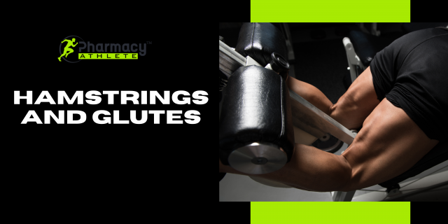
The hamstring muscles are located on the back of the thigh while the gluteus maximus muscle are located at the buttocks. The hamstrings include the biceps femoris, the semitendinosus and the semimembranosus muscles.
They are involved in flexing the knee, but they also work with the glutes in extending the legs at the hips. The glutes, on the other hand, are involved in extending the legs back and kicking the legs behind the body.
The calves also refer to the two separate muscles on the lower leg—the gastrocnemius and the soleus—which is responsible for extension at the ankle.
The hamstring and glutes anatomy:
- Biceps femoris: It begins at the ischium of the pelvis and connects to the fibular, which is one of the bones of the lower leg.
- Semitendinosus: It connects the ischium to the tibia.
- Semimembranosus: It also runs from the ischium to the tibia.
- Gluteus maximus: It is attached to several points including the pelvis, sacrum and coccyx.
The calf muscle anatomy:
- Gastrocnemius: It begins at the base of the femur and connects to the heel bone through the Achilles tendon.
- Soleus: It runs from the back of the tibia and connects to the heels.
The hamstring and glutes physiology:
- Biceps femoris: It is responsible for the extension of the hip, flexion of the knee and outward rotation of the lower leg.
- Semitendinosus: It extends the hip, assists in the flexion of the knee and rotates the lower leg inward.
- Semimembranosus: It has the same function as the semitendinosus.
- Gluteus maximus: It contracts to move the leg rearward, straightens the trunk and returns to the upright position after stopping. It also helps to stabilize the upper body when standing.
The calf muscle physiology:
- Gastrocnemius: It flexes the ankle and offers overall stabilization.
- Soleus: It flexes the ankle and offers overall stabilization.
Fitness training tip:
- Traditional quadriceps exercises like the squat, lunge and step-up also work on the hamstring and gluteal muscles.
- The Romanian Deadlift is one of the best hamstring exercises and it also works on the glutes due to hip extension.
- The biceps femoris can be targeted with lying and standing curls while the semitendinosus and semimembranosus are targeted with seated leg curls.
- Standing calf raises focuses on the gastrocnemius while seating calf raises are best at targeting the soleus.
Read my blog on the Black Panther Program Workout Here.